


Varicose veins of the lower extremities are characterized by dilation of the superficial veins of the legs, which is accompanied by impaired blood flow and valve insufficiency. As a result, the vessels increase in length and diameter, acquiring a serpentine, cylindrical or sacral appearance, although there is also a mixed manifestation of the listed deformations.
Features of the venous system
The formation and development of varicose veins is directly related to the venous system of the legs, which consists of the following.
- sapenous vessels: small and large;
- deep-seated veins (lower leg and thigh);
- perforated vessels that are the connecting connection of the previous two systems.
Normally, 90% of the blood is transported to the lower extremities through deep veins, and the remaining 10% through superficial blood vessels. When it returns to the heart, this mechanism is supported by valves in the walls of the arteries. When the next part of the blood arrives, they are pounded to prevent it from moving from top to bottom under the influence of gravity. Muscle contractions allow the blood to flow normally, pushing the blood further into the heart.
When a person stays upright, blood stasis can develop, which increases the pressure in the arteries and increases their diameter. This process leads to incomplete closure of the flap leaflets, resulting in blood flow disturbed by reflux from the heart.
Deep vascular valves are more likely to be affected because they carry the most blood and therefore experience maximum load. To reduce the high blood pressure inside, a portion of the blood is pumped through perforated vessels to those that are initially not intended for large volumes. Such a load on the walls of the vessels causes them to dilate and varicose veins to form.
At the same time, blood enters the deep veins without stopping, but due to dysfunction and the normal functioning of the cap leaflets of the perforated vessels, the blood is redistributed to the superficial vessels. As a result, over time, chronic varicose veins develop, accompanied by painful sensations, edema and trophic ulcers.
Causes of the disease
In the past, one of the main causes of varicose veins was called a hereditary factor, but today this theory has been refuted. Of course, in some families it is possible to watch the frequent manifestations of the disease, but this is more due to the characteristics of life in the family: eating culture, passive rest, sedentary work and so on.
The development of varicose veins is based on the presence of reflux in the venous system when blood circulates inside the veins in the opposite direction. Additional blood transfusions from deep-seated vessels to superficial vessels are possible due to congenital or acquired degenerative pathology in the Ghanaian apparatus. This causes the superficial arteries to become overfilled with blood and constrict when venous nodules form.
One of the main reasons for the development of varicose veins is an unhealthy diet, which in some cases leads to obesity. Such people are less active, eat mostly high-processed foods, and the proportion of plant fiber in the diet is minimized. After all, they are involved in strengthening the walls of blood vessels and blood vessels, and prevent long-term chronic constipation, which greatly increases intra-abdominal pressure and thus provokes varicose veins. It is noted that an increase in body weight of more than 20% increases the risk of disease by five times.
The main motivating factor for women is to carry a child, the risk of varicose veins increases with each subsequent pregnancy. Heavy weight gain and uterine enlargement put a lot of stress on stagnant legs. This condition is aggravated by the constantly increasing intra-abdominal pressure and the effect of the hormone progesterone, which affects the condition of the elastic fibers in the walls of blood vessels.
Other factors that cause varicose veins of the lower extremities include:
- sedentary lifestyle, standing upright during the day (e. g. hairdressers), long flights or long trips. All of this causes stagnant processes in the lower extremities when blood collects in the superficial arteries and is poorly transported to the heart;
- sometimes uncomfortable for women, wearing tight shoes, especially high-heeled models, increases the risk of developing varicose veins;
- corsets and tight underwear compress the inguinal veins and increase intra-abdominal pressure, which is a direct condition of varicose veins;
- high blood pressure;
- smoking, which indirectly causes thinning of the walls of blood vessels.
Classification of the disease
Varicose veins of the lower extremities are classified according to the prevalence, localization of venous lesions, as well as the presence of pathological reflux, which is characterized by impaired blood flow. There are 4 forms of varicose veins:
- intracutaneous and subcutaneous varicose veins (segmental) without pathological flow of venous blood;
- when segmental varicose veins occur through reflux perforation or superficial veins;
- a common form of varicose veins in which perforation and reflux occur through the superficial veins at the same time;
- varicose veins are characterized by reflux in deep veins.
After varicose veins of the lower extremities become chronic, phlebology considers three degrees:
- Transient edema that occurs periodically on the background of "heavy legs" syndrome.
- Persistent, persistent edema. Hyperpigmentation and eczema may appear.
- Venous ulcer of a trophic nature.
The latter is the most difficult to treat, as it requires early removal of inflammation and healing of skin tissues.
Stages and symptoms
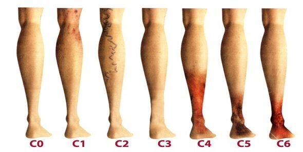
The disease develops so slowly that the symptoms make the patient seek the advice of a phlebologist, sometimes lasting more than a decade. Early manifestations of varicose veins are often associated with fatigue, age or other causes. To fully review the symptoms of the disease, the manifestations are classified according to the stages of varicose veins:
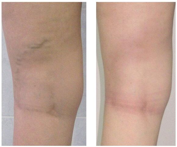
- The first stage begins to manifest itself more often at a young age - after 20 years, when there is a feeling of heaviness in the legs, edema may appear, which completely disappears overnight. Inside the lower leg, you can see an enlarged vein showing itself with a round bulge of skin. At this stage, many people see small spider veins. In general, the symptomatology is subtle and rarely attracts the attention it deserves.
- The second stage is characterized by an increase in the external manifestation of the dilated vessel. The disease already develops against the background of pathological work of venous valves, so the saphenous veins increase significantly in size, and their elongation can also be noted. More often there is heaviness and burning in the legs, they get tired quickly with long walks.
- The disease is already chronic due to persistent imbalance outside the venous blood. In the evenings, patients suffer from edema near the ankle, which can be very intense. There is heaviness in the legs and there may be cramps at night.
- In the absence of treatment in the early stages, chronic failure of the venous system has a negative impact on metabolic processes in the skin, especially in the lower part of the foot. Darkening of the skin near the ankle - hyperpigmentation, thickening and inflammation over time. The condition described is called lipodermatosclerosis. If you do not start venous therapy at this time, trophic ulcers will soon begin to form.
- The fifth stage is accompanied by numerous trophic ulcers, some of which heal periodically with the formation of scars.
- Extensive ulcers open in the area of long-standing trophic disturbances. This condition requires urgent active therapy aimed at both the treatment of varicose veins and the healing of skin ulcers.
Diagnostics
External examination of the lower extremities, palpation of the vessels and preliminary assessment of the stage of the disease are performed in the vertical and horizontal positions of the body. The patient is sent for a general blood test, which allows to study the picture of the disease more accurately:
- the tendency to thrombosis will be reflected at the platelet level;
- hemoglobin level and red blood cell count indicate the degree of blood clotting;
- Due to the increased levels of leukocytes, inflammation can be judged, which helps to diagnose thrombophlebitis more quickly.
Be sure to examine the venous system of the legs, where there are many methods:
- ultrasound dopplerography - USDG;
- phlebography;
- CT phlebography;
- duplex angiocanalization - USAS;
- phleboscintiography;
- photoplepsography;
- phlebomanometry and the like.
In practice, patients are often prescribed USAS and USG, as it helps to fully study the venous system of the legs and identify degenerative areas. The remaining methods can be additionally prescribed if the ultrasound examination does not give a complete picture of the disease. Some of these methods may include complications such as venous thrombosis, catheter perforation of the vessel wall, and allergy to a contrast agent. Consider the most commonly used techniques in phlebology:
- USAS allows the assessment of anatomical, hemodynamic and functional pathologies of the venous bed. The information obtained is subject to computer processing, after which a model of the venous system can be viewed on video or printed on paper.
- Doppler ultrasound determines the permeability of superficial and deep vessels, blood flow velocity with high accuracy. Doppler ultrasound allows you to assess the performance of the valve.
After extensive diagnosis, the doctor prepares a patient's phlebocardium, which allows to determine the damaged segments, degrees and length of the venous system. After that, an appropriate treatment is selected.
Treatment
It is carried out comprehensively and is determined based on symptoms, stage of the disease and the results of the study. In the initial stages, conservative therapy consisting of the following is prescribed.
- When a group of drugs is prescribed drug treatment:
- antioprotectors and phlebotonics;
- anticoagulants;
- disagreement
- local preparations (ointments, gels);
- anti-inflammatory drugs.
- Elastic compression (rarely) where compression stockings or bandages are used. Allows you to give a dose of muscle contraction, prevents stagnant processes, improves blood flow through the arteries. When wearing such underwear, there is an effect that artificially protects the vascular tone.
- Among physiotherapeutic methods, the best treatment results are shown by electrophoresis, diadynamic currents, laser radiation and magnetic field.
- Physical activity that can only be performed in compression underwear (excluding swimming). Cycling, swimming, jogging are recommended. The phlebologist selects a series of individual exercises for the lower extremities that will train the veins of the legs every day.
In addition, patients are advised to take contradictory five-minute procedures every evening in the shower, alternating between hot and cold water. Such manipulations improve blood flow and tone blood vessels.
At the beginning of treatment, it is important to identify the trigger factor in order to have an effective effect on the disease. And patients at risk should visit a phlebologist every 2 years for a prophylactic examination and ultrasound examination of the vessels in the legs.
When conservative treatment does not work or varicose veins are observed at an advanced stage, surgery is used. Today, varicose veins can be completely treated with the following methods:
- Phlebectomy. The essence of the operation is to remove the main stems of the superficial vessel to eliminate the pathological discharge of blood. Perforated vessels are often closed for the same purpose.
- Sclerotherapy. It consists of the insertion of a sclerosis into the affected area of the vessel, which leads to the fusion of the walls. Recently, due to technology, foam sclerosants have been actively used for the same purposes. Blood flow from the defective area is stopped and the cosmetic defect in the form of protruding nodules is eliminated. After such an intervention, no wounds remain, all manipulations are performed in an outpatient setting without further inpatient care. However, sclerotherapy is used only for the combination of small branches of venous trunks.
- Laser coagulation. With the help of a laser beam, the marked part of the vessel is heated, the walls stick together and the blood flow through it stops. However, this technique is only indicated for vessels with a dilation diameter of less than a centimeter.
Prevention
Prophylactic measures can be aimed at preventing the development of both primary and varicose veins, and can be secondary when required to reduce the risk of postoperative relapse or to prevent the progression of the disease. Useful tips:
- lead an active lifestyle without heavy loads on your feet: swimming, walking, cycling;
- look at your weight;
- lift both legs more often;
- Do not wear tight underwear and heels higher than 4 cm;
- use orthopedic padding;
- take a contrasting shower;
- do preventive leg exercises for five minutes daily;
- Wear compression stockings for long walks.
If you see the slightest suspicion of varicose veins - prominent nodules in the legs, swelling, heaviness, do not delay your visit to the phlebologist. Indeed, over time, this insidious disease can lead to many complications, including thrombophlebitis and thrombosis.